A Whole New World!
New Science Labs InsPire STEM Students and Intrigue Others
Timothy Russell, ’12
 The Payden Academic Center’s 11 light-filled laboratories have been an incredible boon to Trinity’s science community – students and faculty alike. Bursting with the latest scientific equipment and classroom technologies, the Payden Center supports cutting-edge research collaborations, while providing an inviting space to learn and study.
Trinity’s scientists see the Payden Center as huge boost to their enthusiasm and dedication to scientific pursuits. When biochemistry major Kimberly Cruz ’18 entered the Payden Academic Center on the first day of her junior year, the difference from her previous science classes was profound. “When I walked in and saw the Trinity seal in the middle of the lobby floor, it really hit me – wow, this is our building, this is really here and I’m really breathing the air – it even smelled different.” For months, Cruz had been following the construction of the building with interest, even checking progress on the live web cam. “It really showed me how much science is appreciated here; the building itself is recognition of how much the scientific community can contribute to Trinity, and to society in general.”
The Latest Equipment
Cruz enjoys working in the new space, and relishes the new equipment available to help her with her research. “Now we can do cell cultures, which is amazing. In order to grow certain bacteria, you need a sterile environment. In this new building we have hoods where we can sterilize everything and grow a competent bacteria to reproduce certain experiments. Over the summer, I did research at Children’s National Medical Center on multiple sclerosis using cultures of oligodendroglioma cells – cancer cells in the brain. To reproduce this experiment here at Trinity, we needed these hoods as well as incubators to grow the bacteria. I could not have done it in the old science building.” Dr. Cynthia DeBoy, Cruz’s advisor, helped her design a summer research experiment specifically to take advantage of the new features and technologies in the Payden Center, allowing Cruz to continue her significant research experience with one-on-one faculty guidance on Trinity’s campus.
Cruz also points out the new sterilizer technology that she uses every day. “We don’t have to use an open flame to sterilize our glass pipettes; we just stick them into the slot of the sterilizer and this blue light sterilizes them. It’s a lot safer to perform these experiments in the enclosed space of the hood. Whereas before there was a Bunsen burner to one side, and the vacuum going on the other side, and serums over there – it is very meticulous work to begin with. Now we just stick the pipette in the sterilizer and take it back out and that’s it.” She no longer has to worry about burning herself, noting “nothing catches on fire when I’m already dealing with bacteria or corrosive chemicals!”
Cruz’s research has given her, and Trinity, recognition on a national level. “I’ve had incredible opportunities because of the research I’ve performed,” she proclaims. At the University of Maryland, Baltimore County, National Symposium, Cruz won first place in the biochemical and physical sciences division, and at another conference in Tampa, she was chosen to give an oral presentation – an honor awarded only to the top 10 abstracts submitted from across the entire country. “It was a really big deal, and I was very nervous,” she recalls with a laugh, “but everyone was very supportive and it turns out I felt like I was really in my element among all these other scientists.” This summer, Cruz will continue to do research as an Amgen Scholar at the prestigious Summer Research Program at Stanford University.
A Space to be Engaged
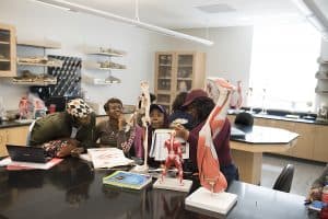
The very architecture of the classrooms and labs has revolutionized scientific activity at Trinity – even drawing in students from other disciplines with impressive displays of science in action. Clare Booth Luce Assitant Professor of Biology Mia Ray has observed that, “The anatomy lab has this glass wall where people can look in, and people walking past the lab stop and stare: ‘What are they doing in there?’ This happened just yesterday when my students were using clay to build muscles onto the frame of the skeleton. Other times, I have the microscope image projected up onto the screen and students getting out of other classes who may have never considered science stop and watch.”
In addition to capturing the imaginations of science and other students alike, senior Charlene Valdez notes that her science professors, too, have been motivated by the multidisciplinary nature of the Payden Center, offering lessons that go beyond the traditional idea of a science class. She recalls how Clare Boothe Luce Professor of Biology Karobi Moitra recently demonstrated how students can apply scientific knowledge in other disciplines with a project that used evolutionary concepts in the creation of a business model.
Valdez, whose previous work includes research on pancreatic cancer at MIT in the summer of 2016, says, “I wanted to continue my undergraduate research! I have ideas that I want to push forward and develop, and now I can with these updated facilities and technologies in the Payden Center. It just wasn’t possible before.” Valdez’s research and academic accomplishment have earned her a spot this fall in New York University’s graduate program in biology. Her research will focus on bioinformatics and systems biology.
Classroom Technology
Like Valdez, Assistant Professor Ray loves the cutting-edge technology integrated into every Payden learning space, and makes use of new digital projectors in almost every class she teaches. Virtually any information or demonstration can be shown on large screens in every classroom and lab in the new building. The limitless options provide a much richer, visually augmented way to present material. With a screen that can display real-time information from all types of scientific devices, Ray can show her biology classes exactly what cells and features to look for by projecting her own microscope’s view for all to see. The students get a better idea of “what they are supposed to be looking at – it gives them more guidance. Not everybody looks into a microscope the same way.” After demonstrating how to adjust the microscope, Ray clicks over to a PowerPoint presentation that explains what the students are looking for, then plays a quick video of live cells.
The students’ microscopes are also technologically advanced, and can capture images of cells and other structures, which students then manipulate – sometimes for their lab reports, and sometimes for more aesthetic purposes. Trinity’s campus science publication, Metamorphosis, features artworks created from these captured images. (See sidebar.)
Junior biochemistry major Raissa Audrey Tseumie uses the microscopes in her genetics class to examine cells as part of a group project. While she operates the microscope, the rest of her group use an app called Leica AirLab on their mobile devices to see what she is looking at in real time. Tseumie was recently inducted into the Phi Beta Kappa honor society, and will be continuing her research over the summer at the University of Virginia’s Summer Research Internship Program.
Like Tseumie, all of Trinity’s science students and faculty find themselves and their research reinvigorated with new equipment, beautiful spaces, and the latest technology to support them.